Industrial X-Ray & CT of Metal Foam – Helical
Helical CT Scanning for Oblong Parts
We receive many oblong parts for X-ray/ CT inspection. From 3D printed tubes, to medical parts and assemblies, and lightened foams and composites. Helical scanning is important because it allows us to simultaneously reduce noise and maintain high resolution for intricate, high detailed objects. Traditional circular scanning resolution is limited by the object to source distance, so oblong parts would need to be staged with consideration of their largest dimensions.
Helical allows us to increase magnification and corkscrew the part from top to bottom, ignoring an object’s overall height. The staging and imaging limiter becomes the largest width rather than height.
This case study reviews extremely versatile metal foams used for a variety of sectors including aerospace, filtration, shielding, and biomedical. The largest sample, 10x1x1 inches, was scanned at just 20 micron voxel resolution. The raw dataset was 186gb! All data is shared with permission.
Slice Data
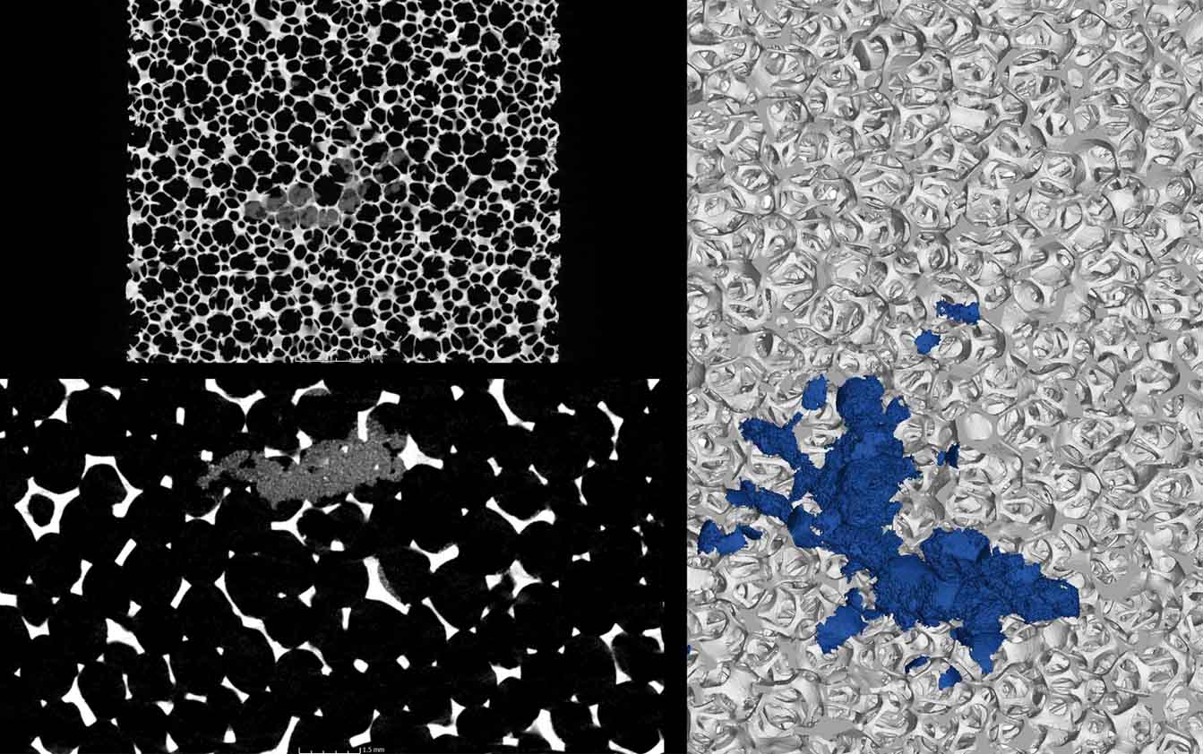
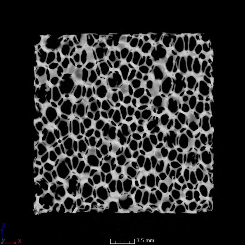
The below images are slice views through the reconstructed CT data set. For this application, abnormally large cell or pore sizes, inclusions, and other defects are detected and imaged.
The first image is a single slice; the second image is a slice showing standard deviation from a 3mm zone.
Imaging Filters & Techniques
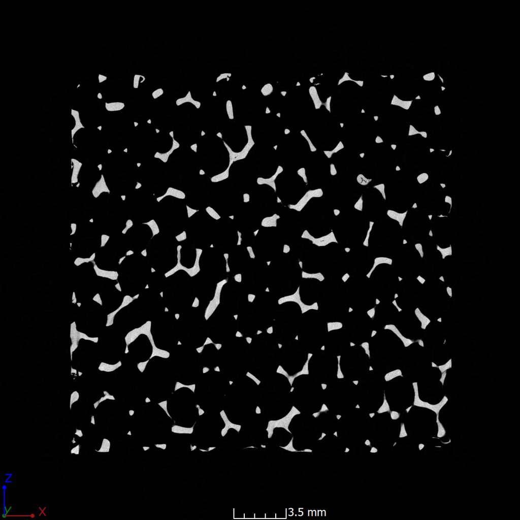
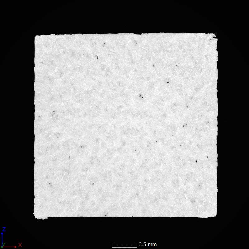
Applying filters to the CT slice data allows us to investigate certain requirements. For example, the below image has a 30mm maximum gray value filter applied. This shows the highest gray values in a single slice from 30mm of data, and allows us to find long flow, leak, or structural paths.
Slice Clip Reporting Method
Slice data can be exported into clips so the entire part can be analyzed and saved in a manageable format. The clip can be paused so that specific locations can be saved as images.


Defect Detection and Analysis
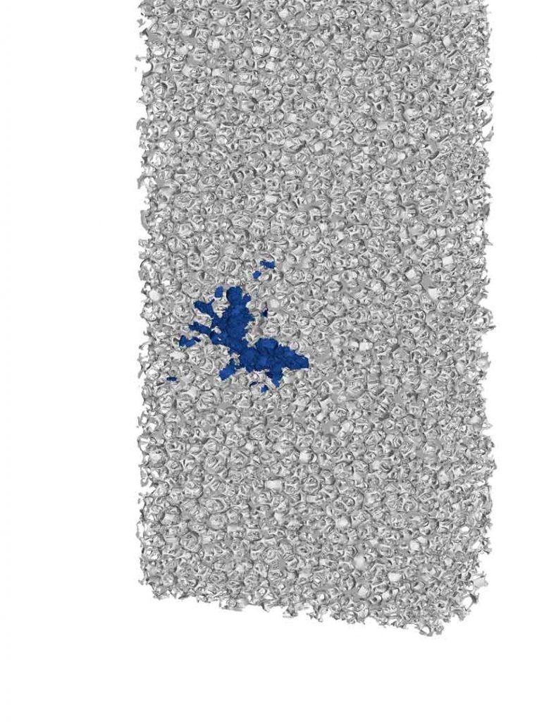
This image shows an inclusion within the metal foam. The inclusion was extracted and rendered separately. Volume and location can be reported. The scan data can also be exported into .stl or other formats to be analyzed in other softwares.
