Detecting and Quantifying Part Deformations using Industrial CT Scanning
Detecting Part Changes using Industrial CT Scanning
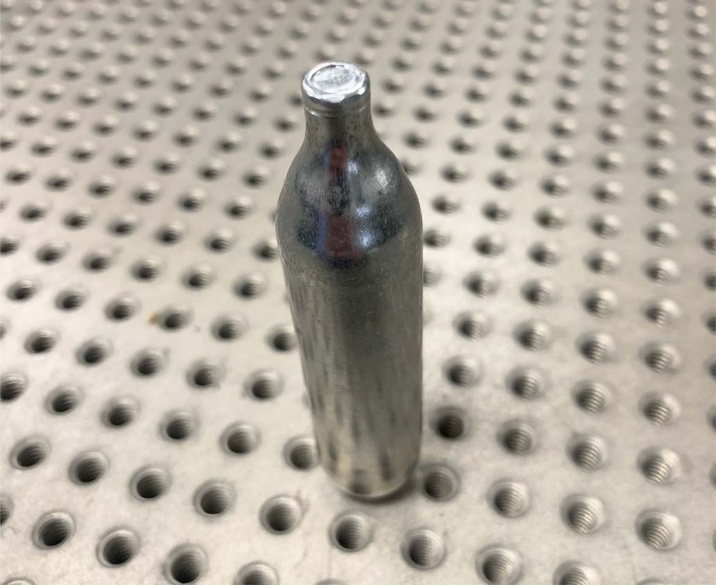
High Resolution Industrial CT scanning allows for the detection of extremely small part deviations. Because an entire part can be rendered and visualized, small changes in shape can quickly be quantified for various analyses. In this case study we demonstrate the change in profile after a pressurized CO2 cartridge has been evacuated. Applications for this analysis could be for wear, stress, or use studies to fully understand how a part or assembly changes.

Testing Repeatability of Method
To detect the change in profile between a pre-to-post pressurized CO2 cartridge we must first understand the repeatability of the measurement method. To do this we scanned the same part twice, but replicated the actions as if it were a pre & post scan. This means we used the same scanning parameters, but removed and re-loaded the sample into the scanning position.
The results are incredible. The image shows a profile deviation scale of +- 10 microns. 99%+ of the surface profile between scan 1 (Gray) and scan 2 (Blue) is below 2 microns. There are some outlier locations that show 4-7 microns.

Quantifying Deformation
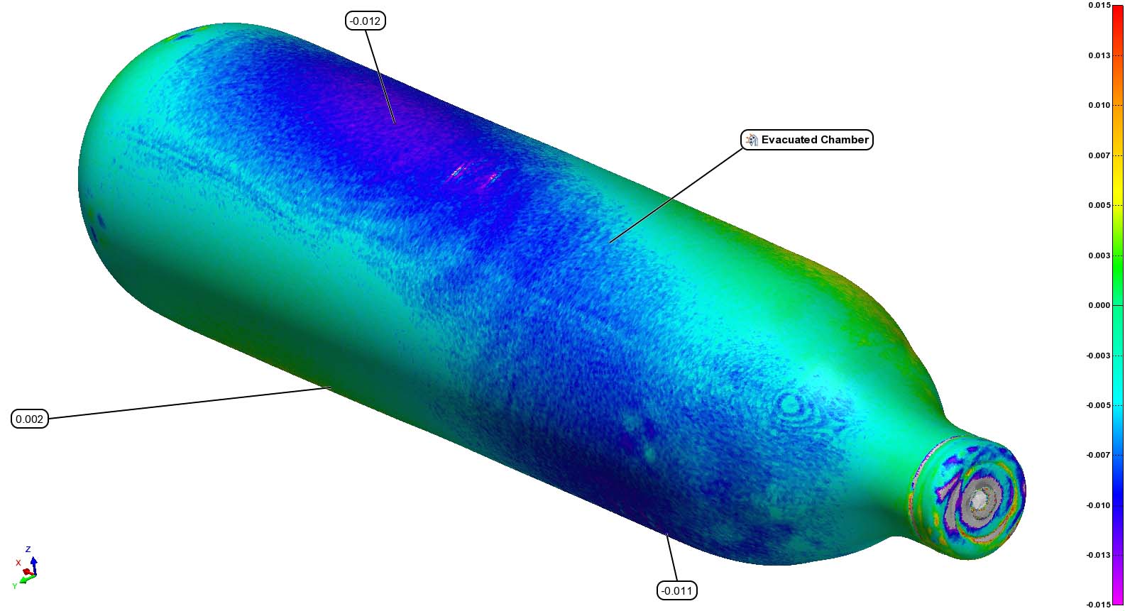
With an understanding of our average repeatability for this scenario, we depressurized the CO2 cartridge, re-loaded the sample into the system, and scanned using the same parameters. After exporting the new scan as mesh from Volume Graphics we applied the same alignment and overlay in PolyWorks.
While the overall deformation is quite small (10-12 microns) it shows that we can quantify with certainty for a given sample. Insights from the heatmap show trajectory of deformation, where areas of interest are, and associated tangible profile values. Repeating this process across many samples would show trends that could lead to value added process changes.

Detection of Inclusions or Fragments using CT Slice Data
The below images show slice views of the interior of the cartridge which contains free fragments and a 3D rendering of the same. These fragments were present before depressurization


